Author:
Monica Porter
Date Of Creation:
18 March 2021
Update Date:
1 July 2024

Content
Macular degeneration or age-related macular degeneration (AMD) is the leading cause of vision loss in people age 60 and older. This is a painless condition that affects the macula - part of the retina that focuses on the central visual area. The yellow point is also the part that helps you read, drive a car, recognize people's faces and other images. Although there is currently no cure for macular degeneration, you can alleviate its effects through lifestyle changes, eye care therapies, and other preventive measures.
Steps
Part 1 of 5: Understanding the disease
Learn about the stages of AMD. Your ophthalmologist will determine which stage of AMD you are at based on the amount of drunsen in your eye. Drunsen are yellow or white deposits that accumulate in the retina.
- Early stage: the drusen is about the size of a medium to the diameter of the hair and does not lose vision.
- Middle stage: large drusen and / or change in pigmentation; Usually no vision loss.
- Late stage: This stage has two forms:
- Dry macular degeneration: The photoreceptors of the macular are damaged. The eyes cannot use light to transmit images to the brain. You may gradually develop an attack and lose your vision.
- Wet macular degeneration: This condition is caused by abnormal growth of blood vessels, gradually swollen and ruptured blood vessels. Fluid builds up inside and below the macula and changes vision. The disease progresses more quickly than the dry macular degeneration.

Understand the development of "dry" macular degeneration. Dry macular degeneration occurs due to degeneration of the cells in the retina. The condition that cells degenerate or die, plus lack of fluid is the reason why the disease is called "dry" macular degeneration. These cells are also called photoreceptors, which are cells that use light entering the retina to help the brain understand images through the cortex in charge of the visual organ. Basically, the light-sensitive areas help us to understand what we are looking at.- As we age, the accumulation of fatty deposits called drunen in the macula leads to degeneration. During eye examination, accumulation of drusen is detected as yellow dots on the macula. AMD does not lead to total blindness, but it significantly limits the central visual area.
- The "dry" macular degeneration is more common than the "wet" form. Dry macular degeneration has the following signs and symptoms:
- The image of printed text is blurred.
- Need more light when reading.
- Difficult to see in the dark.
- It is difficult to recognize faces.
- The central visual area is significantly narrowed.
- Blind spots are clearly visible in sight.
- Gradually decreased vision.
- Confused geometrical images or still lifes.

Know about "wet" macular degeneration. This form of AMD occurs when abnormal blood vessels grow below the macula. Due to the larger macular size, blood vessels can leak fluid and blood into the retina and macula; In rare cases, complete perforation of the retina and macula. Wet macular degeneration is more rare than dry form and is a more serious eye disease that can lead to blindness. The cause of wet macular degeneration is unknown, but many studies have shown risk factors for developing the disease as you get older. Signs and symptoms include:- Look at the lines in a wavy shape.
- Blind spot appearance.
- Loss of vision in the central region.
- Quickly lose vision.
- No pain.
- Scars form in the blood vessels, which can lead to permanent vision loss if not treated promptly.
Part 2 of 5: Know the risk of developing the disease

Awareness of the aging process. Macular degeneration is an age-related disease. The risk of developing AMD increases with age. At least one-third of the elderly over 75 have some degree of AMD.
Understand that genetics play an important role. If you have one parent or both have AMD, you will also be more likely to develop AMD by the time you turn 60. Don't forget, however, that genetic factors aren't everything, and how to take care of yourself is important equally important.
- In general, women and whites are at a higher risk of developing AMD.
Know that smoke is a high risk factor. Smokers are at an increased risk of developing this condition. Numerous studies have demonstrated a link between smoking and macular degeneration. Tobacco smoke also damages the retina.
- If you are a smoker (especially women or whites), you should be on the lookout for macular degeneration, even if symptoms don't appear.
Track health status. Your overall well-being may indicate a risk factor for developing AMD. People with high blood pressure or diabetes are at high risk.
- Even people without diabetes but on a high-carb diet with a high glycemic index tend to develop macular degeneration as they age. Remember, blood oozing from the blood vessels of the retina is a sign of wet macular degeneration. This is worse when the arteries become blocked due to the buildup of plaque.
Consider your surroundings. How often are you exposed to fluorescent light? Ultraviolet light from fluorescent light is believed to increase the risk of eye diseases. Additionally, the level of risk may increase if you live in a sunny area and your eyes are often exposed to the sun. advertisement
Part 3 of 5: Getting medical treatment
See an ophthalmologist. The ophthalmologist will diagnose the disease during routine eye exams. Your doctor will use eye drops to dilate the pupils in the eyes. In the case of dry macular degeneration, the doctor can easily detect drusen during an examination.
Test eyes with Amsler grid (Amsler grid). You will be asked to look at the Amsler grid, a chart-like table. If you see some wavy lines, you may have macular degeneration. To check for symptoms, you can print the Amsler grid on the Prevent Blindness website and follow these instructions:
- Place the chart in line of sight, 61 cm away from the eye.
- Put on reading glasses and cover one eye with your hand.
- Focus on the midpoint for one minute, repeating steps with the other eye.
- If any lines appear wavy, you should contact an eye care professional promptly.
Ask your ophthalmologist about an ocular angiogram. To do this procedure, a dye is injected into a vein in your arm. The dye is then taken as the dye travels up the veins in the retina. This method can detect a leak, a hallmark of wet macular degeneration.,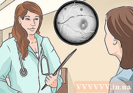
- 8-12 seconds after injection, the dye should be seen on the optic nerve.
- 11-18 seconds after injection, the dye should be seen in the retinal area.
Optical tomography (OCT). This method uses light waves to look at multiple layers in the retina. This test can assess the thickness of the retina, the structure of the retinal layers, and any abnormalities in the retina such as fluid, blood or new blood vessels, if any.
- You may have dilated pupils in your eye prior to the OCT scan, although OCT is also likely to work through the non-dilated pupil.
- Next, you will place your chin on the chin rest to keep your head steady and inactive.
- A ray of light will shine into the eye.
- Using light waves, this method can detect living tissue within seconds and painlessly.
Consider injecting anti-VEGF agents. Vascular endothelial growth factor (VEGF) is the primary chemical that causes the abnormal growth of blood vessels. When this chemical is inhibited through antiangiogenics, also known as antiangiogenics, the growth of blood vessels can be inhibited. Your doctor will determine if this option is right for you.
- Bevacizumab is a new popular anti-vascular agent. The usual dose is to inject 1.25 to 2.5 milligrams of drug into a vitreous cavity in the eye. Usually this drug every 4 weeks is injected once, for 4 to 6 weeks. Other drugs like Ranibizumab have a dosage of 0.5 mg, and Aflibercept 2 mg.
- The procedure will be done using a very fine needle and local anesthetic to relieve pain. In general, the whole procedure should be painless and only slightly uncomfortable.
- Side effects can include increased pressure in the eye, infection, bleeding, and damage to the lens.
- You should have better eyesight within a year, improvement can be noticed after two weeks and usually peak in the third month after the third injection.
Explore photodynamic therapy (PDT). This is light therapy and a drug to stop blood vessel growth. This therapy may be only effective in treating wet macular degeneration.
- This is a two-step procedure done in one visit. A substance called verteporfin or visudyne will be injected into a vein. This drug works to suppress blood vessel growth, which occurs in wet macular degeneration, and is taken 15 minutes prior to photodynamic therapy.
- Then, light of the right wavelength is shone in the eye, focusing on the abnormal blood vessels. The light will activate the verteporfin that was previously injected to block the leaking blood vessels.
- Light is adjusted with the appropriate wavelength, eliminating the risk of scar tissue causing vision impairment.
- Check with your doctor to see if this method is safe for you. Anti-VEGF is currently the standard treatment of choice of first choice, and PDT is sometimes used in combination with Anti-VEGF.
Seek immediate medical attention when severe symptoms appear. Go to the nearest emergency care facility or contact an ophthalmologist immediately if you experience sudden headache, change in vision, or any unexplained pain during treatment. advertisement
Part 4 of 5: Using vision aids
Use a magnifying glass. When macular degeneration occurs, the central visual area is the most affected, while the peripheral visual area remains somewhat unaffected. Hence, people with macular degeneration can still compensate for their peripheral vision. Magnifying glass works to magnify objects, helping the patient to see more clearly.
- Magnifying glass has magnification from 1.5 to 20 times. You can easily take your magnifying glass with you thanks to its compact size. Many foldable types are available in pocket size.
- Try a stand magnifying glass. These typically have 2 to 20 magnifications and can be erected, so you don't have to hold them with your hands. This type of magnifying glass is very useful for patients with hand tremors. Some magnifying glasses have a base with additional lights for use in low light.
Use monocular or a telescope. This device has a magnification of 2.5 to 10 times, and comes in handy at a distance.
Use binoculars. This device has the same type of magnification as a telescope, and you can use both eyes to see objects.
Use a magnifying glass. This type of magnifying glass is attached to the patient's eyeglasses and helps to see far away. The eyeglasses magnifying glass allows the patient to change between long-range vision and telescope field of view. There are also eyeglasses for normal viewing.
- These glasses work similarly to bifocals.
- These glasses are recognized and prescribed by a vision therapist.
Use TV magnifier. This is a TV camera with a kickstand that magnifies the writing on the video screen. You can use this type of magnifier to aid in a variety of tasks such as reading, writing and viewing photos. Some devices may even underline or highlight information. This type of device can be used with a computer.
Use a reader to make a sound. This machine will read printed text out loud.
- Use character recognition (OCR) software to turn your personal computer into a reader. ,
Learn about absorbent lenses. These lenses work by absorbing light transmitted through the eyes, helping to reduce glare and harmful ultraviolet rays.
- These lenses can switch between bright and dark areas.
- These lenses can be used on prescription eyeglasses.
Part 5 of 5: Eye Care
Get regular eye exams. Although not preventable due to age factors, macular degeneration can be detected early and treated promptly with regular eye exams. Early detection of macular degeneration can help slow vision loss.
- Starting at age 40, you should have routine eye exams at least every six months or as recommended by your ophthalmologist.
Ask your doctor about special eye exams. You should have your ophthalmologist perform eye exams to detect drunsen, damage to your blood vessels, change in pigmentation in the retina and visual disturbances. Some of the tests that detect vision disorders are:
- Visual acuity test: This test uses graphs to check vision from a distance.
- Amsler grid: This type of test checks for central vision disturbances by showing the patient whether the lines are straight or wavy. If the lines are wavy, the person may have macular degeneration.
- Dilated eye exam: During this test, the pupils in the eye are dilated so the doctor looks at the optic nerve and retina to assess damage. Your doctor will also check for pigmentation changes in the retina. Pigments that appear in the retina indicate poor light reception.
- Fluorescence retinal angiography: This exam will evaluate the arteries in the eye to detect leaky blood vessels. The doctor will inject a dye into a vein in the patient's arm.
- Optical tomography: The exam is done after the pupil is dilated. The infrared light is then used to scan the retina, through which the doctor can identify damaged areas.
Avoid smoking. In addition to its other harmful effects on overall health, smoking also leads to macular degeneration. Smoking plastic can stimulate the formation of drusen (waste products that accumulate in the eye). In addition, tobacco also contains caffeine, which is considered a stimulant that can raise blood pressure. The blood vessels under the retina and macula can easily rupture when blood pressure is high.
- Smokers are twice as likely to have macular degeneration than nonsmokers. Tobacco is bad for you, your eyes and other organs in your body, and even those around you.
- Even if you stop smoking, the effects may remain for a few years. Consider this as an excuse to start your smoking cessation journey as early as possible.
Take control of pre-existing conditions such as high blood pressure. Take medicine, get regular medical check-ups and make lifestyle changes to adapt to your health condition.
- For example, if you have high blood pressure and are diagnosed with wet macular degeneration, the damaged blood vessels in your eye will find it difficult to recover due to increased blood pressure. This can increase the risk of blood vessel rupture, leading to more blood leakage.
Exercise regularly. Physical exercise has many health benefits, including eye health. Drusen formation is associated with high levels of cholesterol and fat. Exercise can burn fat and eliminate bad cholesterol, preventing the buildup of this waste.
- It is recommended to exercise at least three times a week. Focus on aerobic exercises that help you sweat and burn fat.
Add vitamins. The eyes are constantly exposed to intense ultraviolet (UV) rays from sunlight and pollutants from smog. Frequent exposure to harmful factors leads to oxidative damage. Oxidized eye cells can lead to macular degeneration and other eye diseases.To combat this process, you need to eat foods rich in antioxidants. The most common antioxidants are vitamin C, vitamin E, zinc, lutein, and copper.
- Vitamin C: The recommended daily dose of vitamin C is 500 milligrams. Good sources of vitamin C are: broccoli, cantaloupe, cauliflower, guava, bell peppers, grapes, oranges, berries, litchi, and squash.
- Vitamin E: The recommended daily dose of vitamin E is 400 milligrams. Good sources of vitamin E include: almonds, sunflower seeds, wheat embryos, spinach, peanut butter, collard greens, avocado, mangoes, hazelnuts, and rainbow chard.
- Zinc: The recommended daily dose of zinc is 25 milligrams. Some good sources of zinc are: lean beef, skinless chicken, lean lamb, pumpkin seeds, yogurt, soybeans, peanuts, legumes, sunflower butter, pecans, kale, vegetables. Spinach, beet leaves, lettuce, asparagus, okra, coriander, watercress, persimmons, and green beans.
- Copper, lutein and zeaxanthin: Both lutein and zeaxanthin are found in the retina and lens. They are natural antioxidants, helping to absorb harmful UV rays from the sun. Both substances are found in green leafy vegetables.
- Supplement 2 mg of copper per day.
- Get 10 mg of lutein per day.
- Get 2 mg of zeaxanthin per day.
Reduce the amount of beta carotene. Research has shown that beta carotene can increase the risk of lung cancer, especially when the patient is a smoker. The study also showed that beta carotene was ineffective in inhibiting the progression of AMD. Currently, doctors often make a list of supplements that do not contain beta carotene.
Use eye protection, including sunglasses. High levels of UV exposure from the sun can damage the eyes and contribute to the development of macular degeneration. Wear sunglasses that provide UV and blue light protection for best results.
Be careful with some activities. Some activities at first glance are just daily tasks but now you need to be done with caution. Depending on whether your vision is severe or light, you may need to ask a friend, family member or caregiver to help you with some work. In many situations it is better to ask someone for help instead of acting without thinking of potentially harmful consequences. Be careful when participating in the following activities: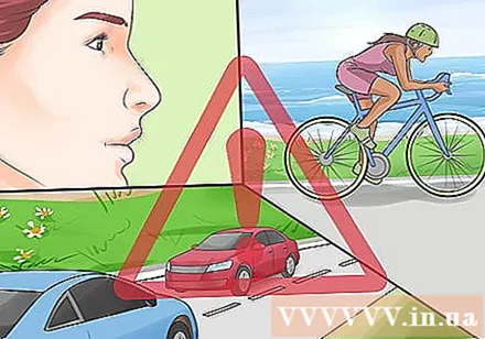
- Driver
- Ride bicycle
- Operating heavy machinery
Information understanding. With macular degeneration, it may feel like your life is suddenly out of control. However, with the care of an ophthalmologist, there are some things you can do to manage your condition as well. Finding the information is the best way to understand the disease and adhere to the treatment regimen. You can start with research into AMD, treatment options, and new technology to help restore the eye. advertisement
Warning
- The most common risk factors for developing macular degeneration are age, family history, race, body weight, and progression of other illnesses.


