Author:
Eric Farmer
Date Of Creation:
12 March 2021
Update Date:
1 July 2024
Content
An electrocardiogram (ECG) allows you to assess the levels of electrical activity of the heart, which is used to diagnose the condition of the heart. This article will teach you to understand the ECG waveform.
Steps
Method 1 of 2: The Basics
 1 Get a referral on an EKG. This is the simplest and oldest method for diagnosing heart pathologies, but at the same time, one of the most informative and quickest, since the printout of the results is usually obtained immediately after the ECG.
 - When you undergo an ECG for the first time, it can be a little exciting: they smear you with a sticky cold gel, cling to your chest and arms and legs ... All this, however, is not just like that: this way the electrical activity of the heart is monitored, and its results will be printed on paper.
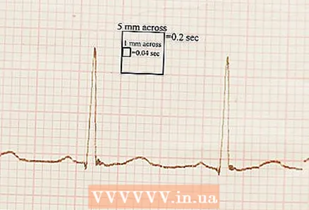 2 What are the cells on the printout for? The vertical axis is voltage, the horizontal axis is time, the large squares are divided into 25 small ones.
 - Small squares have a side of 1 mm and represent 0.04 seconds. Large, respectively, 5 mm and 0.2 seconds.
- A centimeter of height is equal to 1 mV of voltage.
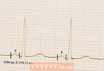 3 Measure the time between heartbeats. This is the so-called. P-wave, a straight line between highs and lows. Its normal length is 0.12-2 seconds, i.e. 3-4 small squares.
 - This value should be the same throughout the ECG. If in one place the P-wave length is one, and in another - another, then this is a sign of an irregular heartbeat. However, if the doctor is calm, then there is no need to worry about this.
- A slight increase after the T-wave, is the end of the heartbeat, means repolarization of the heart valves.
Method 2 of 2: Details
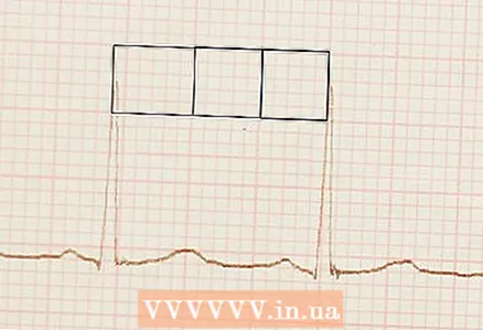 1 Find two similar peaks on the ECG. Count how many squares there are between them. The apex of the peaks is R, and the peak itself is known as the “ventricular complex” or “QRS complex”.
 - The picture above shows normal sinus rhythm, i.e. exactly what will be on the ECG in a person who does not suffer from heart disease. There are, of course, individual characteristics, but in general, a healthy heart works like this.
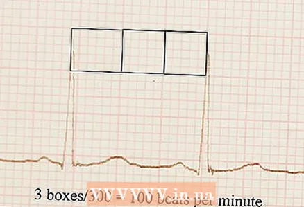 2 Calculate your heart rate using the following formula: Divide 300 by the number of large squares between the two peaks. In this case, the heart rate is 100 beats per minute.
 - If there were 4 large squares, then the heart rate, respectively, would be equal to 75.
- If you cannot find similar peaks on your ECG, count the number of peaks in the 6-second interval and multiply this number by 10. If the ECG shows 7 R-waves in 6 seconds, then the heart rate is 70.
- 3 Ask your doctor about an irregular heartbeat. If you found this on your EKG, and the doctor did not tell you anything, it may be because he simply does not want to bother you over trifles, and not at all because he did not study well and does not know anything.
- If the distance between P and R is too long, it is called "first degree heart block." A “bundle branch block” is when the QRS is longer than 0.12 seconds. Atrial fibrillation - when arrhythmia is superimposed on the absence of direct and long P-waves, instead of which wavy lines are shown on the ECG.
Tips
- The diagnosis based on the ECG should be made by a qualified doctor and only he.


