Author:
Mark Sanchez
Date Of Creation:
5 January 2021
Update Date:
1 July 2024

Content
- Steps
- Method 1 of 2: Preparing for the examination
- Method 2 of 2: Different types of research
- Tips
- Warnings
- Similar articles
An X-ray (also called simply an X-ray) is a painless procedure that allows you to examine the internal organs. With this examination, you can visually separate soft tissue from hard tissue (for example, from bones). X-rays are used to diagnose fractures, bone infections, benign and malignant tumors, arthritis, vascular occlusion, and caries. This research method is also used for digestive problems and if the patient has swallowed a foreign object. Knowing what to expect from your procedure and how to prepare for it will make it easier for you to get through it.
Steps
Method 1 of 2: Preparing for the examination
 1 Talk to your doctor before starting your procedure. It is important to consult a doctor before taking a picture, especially if you are breastfeeding or intending to become pregnant. You will be exposed to small amounts of radiation that can be harmful to a developing fetus.
 - Your doctor may recommend another test.
 2 Ask if you will need to give up food before your procedure. You cannot eat before some tests, but most often this is only required when examining the digestive tract. In this case, you will need not to eat or drink for 8-12 hours before the procedure.
 - If you are constantly taking medication but cannot eat before the x-ray examination, take the tablets with a little water.
 3 Wear comfortable clothes and shoes. Choose comfortable clothes as you will need to take off some things before the procedure and / or sit in line.
 - Wear loose-fitting clothing that allows you to move freely (for example, a button-down shirt; women can wear a bra with a closure in the front).
- If you are about to take a chest X-ray, you will need to strip to the waist. You may be given a special robe.
 4 Remove all jewelry, glasses and metal objects. It is best not to wear jewelry because you will have to remove them before the procedure. If you wear glasses, they will also need to be removed.
  5 Arrive early. You may need to fill out some paperwork, so it is best to arrive early. In addition, the doctor may need to inject a contrast agent prior to the procedure.
 - Be sure to bring your doctor's referral with you. This way the radiologist will know which part of the body you need and how to take it.
- Take your insurance policy with you.
 6 If you need to take a scan of your abdomen, go to the toilet before your procedure. The bladder should be empty. You will not be able to move or leave the office after the procedure begins. Try not to drink a lot of water in the morning.
  7 Be prepared to take a contrast agent if needed. In some studies, a contrast agent is injected, which allows you to better see certain areas in the image. You may be offered:
 - Drink barium or iodine solution.
- Take a pill.
- Get an injection.
 8 Remember that you will need to hold your breath for a few seconds. This will allow you to see the heart and lungs more clearly in the picture. Sometimes you also need to freeze and / or take different poses (it all depends on which organ needs to be examined).
 - The radiologist will place you between the machine and a plate that creates a digital image.
- Sometimes sandbags or pillows are used to hold the body in position.
- You may be asked to change your body position in order to take a multiple-angle shot.
 9 Be prepared for a lack of sensation during the procedure. An x-ray is a painless procedure in which x-rays pass through the body and form an image. The procedure usually takes only a couple of minutes, but if a contrast agent has been used, it may take longer.
Method 2 of 2: Different types of research
 1 Know what to expect from a chest X-ray. This is one of the most common procedures. This image provides an image of the heart, lungs, airways, blood vessels, and bones of the spine and chest. Usually a chest x-ray is used for complaints about:
 - Shortness of breath, severe or chronic cough, chest pain, and trauma.
- A chest scan can diagnose disease and track changes in pneumonia, heart failure, emphysema, lung cancer, fluid or air accumulation around the lungs.
- If your doctor has ordered a chest X-ray, no special preparation is required. Just follow the guidelines above.
- A chest scan takes approximately 15 minutes. Most often the picture is taken in two projections.
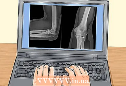
 2 Know what to prepare for when taking a bone scan. X-rays of bones are usually done in cases of fractures and cracks, dislocated joints, injuries, infections, unusual bone growths or abnormal changes in the bones.If you have any pain after your injury, ask your doctor if you can take pain relievers before the procedure, as the radiologist will have to move the bones and joints to take the picture.
 - Bone x-rays are also used to diagnose cancer and other tumors. It detects foreign objects in soft tissue around or inside bones.
- If you are assigned such a study, special training is not required. Follow the guidelines above.
- Typically, this procedure takes 5-10 minutes. Sometimes a scan of the healthy limb is also taken to compare healthy and diseased bones.
 3 Know if you need to take a picture of the upper digestive tract. X-rays of the upper digestive tract are used for trauma and to diagnose diseases of the esophagus, stomach and small intestine. You may also be given an X-ray of your stomach.
 - In this study, a special device is used - a fluoroscope. It allows you to see the internal organs in motion.
- Be prepared to be asked to take a contrast agent prior to the procedure.
- Sometimes, patients are also asked to take baking soda crystals to improve their picture.
- X-rays of the upper digestive tract can determine the cause of swallowing problems, chest and abdominal pain, sour belching, unreasonable vomiting, severe indigestion and blood in the stool.
- This test is used to diagnose ulcers, tumors, hernias, bowel obstruction and inflammation.
- If you are scheduled for a gastrointestinal imaging, you will need to not eat for 8-12 hours before the procedure.
- Remember to go to the bathroom before starting your research.
- This examination usually takes 20 minutes. The procedure may cause bloating and constipation. The stool may turn gray or white and remain so for 48-72 hours after the procedure due to the contrast agent.
 4 Know what to expect from an X-ray of the lower digestive tract. This exam looks at the large intestine, appendix, and sometimes a small area of the small intestine. This type of examination also uses a contrast agent and a fluoroscope.
 - This test is often prescribed for symptoms such as diarrhea, bloody stools, constipation, unexplained weight loss, bleeding, and abdominal pain.
- X-rays of the lower digestive tract are used if benign tumors, cancer, inflammatory bowel disease, or large bowel obstruction are suspected.
- If you are assigned this study, you will need to skip eating in the evening and drink only clear liquids: juice, tea, black coffee, cola or broth.
- You may be asked to take a laxative in the evening to cleanse your bowels.
- Remember to go to the bathroom before starting your procedure.
- The research will take 30-60 minutes. You may feel pressure in your abdomen and mild cramping. After the test, you will be given a laxative to remove the barium from your body.
 5 Find out what happens with X-rays of the joints. Arthrography is a special type of X-ray examination that is used to diagnose joint diseases. There are two types of such research: direct and indirect.
 - In indirect arthrography, a contrast agent is injected into the bloodstream.
- In direct arthrography, a contrast agent is injected into the joint.
- The procedure allows you to find abnormalities in the form of joints, determine the cause of pain or discomfort in the joints.
- Arthrography can also be done with a CT scanner or MRI machine.
- If you need to take this test, no special preparation is required. Follow the guidelines we provide above.
- In some cases, it is necessary not to eat before the procedure (that is, if it is carried out with sedation).
- Arthrography usually takes half an hour. You will feel a slight prick and a burning sensation if you are given anesthesia.
- You may also feel pressure or pain when the needle is inserted into the joint.
Tips
- Ask your doctor or radiologist to tell you what to do before, during, and after your procedure.
- Talk to your pediatrician about how you can help your baby being tested. Parents are often allowed to be present during this procedure.
Warnings
- Tell your doctor or radiologist that you are or may be pregnant.
- Routine X-ray examinations are considered safe, but many doctors do not recommend taking them more than once every six months, and sometimes even once a year, since the examination is associated with exposure to radiation. Sometimes the pictures need to be taken more often (for example, after treatment for pneumonia or fractures). If you are worried about possible exposure to radiation, discuss this with your doctor before starting your procedure.
Similar articles
- How to lower high creatinine levels
- How to check if you have a hernia
- How to get your voice back
- How to get rid of boils
- How to check if a wound is inflamed
- How to reduce muscle lactic acid production
- How to remove swelling from fingers
- How to diagnose a ruptured calf muscle
- How to quickly lose your voice
- How to get rid of pinworms


