Author:
Roger Morrison
Date Of Creation:
17 September 2021
Update Date:
1 July 2024

Content
Prokaryotes and eukaryotes are terms used to refer to types of organisms. The main difference between the two is the presence or absence of a "real" nucleus: eukaryotes have one, prokaryotes do not. While this is the most easily recognizable difference, there are other important differences between the two organisms that can be observed under a microscope.
To step
Part 1 of 2: Using a microscope
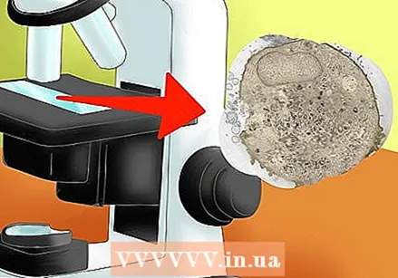
 Use a microscope slide. Prokaryote and eukaryote slides are available from specialist suppliers.
 - If you are in school, ask your physics teacher if he knows how to get slides.
 Place your microscope slide on the microscope table (the platform on which the slides rest). Some microscopes have clips that hold the slide in place to prevent it from shifting during focusing and viewing. If there are clips on the table, gently push the slide underneath to secure it. If there are no clips, place the slide directly under the lens.
 - Be careful when sliding the slides under the clips. Too much force can damage the slide.
- You may have to move the slide while looking through the eyepiece to find the desired area of the specimen.
 Make sure the microscope is at the lowest magnification. The part of the microscope that allows magnification is called an objective. Compound light microscope objectives usually range from 4x to 40x. You can go up to higher magnifications if needed, but if you start low you can easily find the specimen on the slide.
 - You can determine the magnification of the lens by looking at the lens itself (it has a label).
- The lens with the lowest magnification will also be the shortest, while the lens with the highest magnification will be the longest.
 Focus the image. Looking at a blurred image makes it difficult to distinguish small structures and define aspects of the cell. To see every detail more clearly, make sure the image is in focus.
 - While looking into the eyepiece, use the focus buttons located under the object table on the side of the microscope.
- By turning the knobs, you can see the image become sharper or less sharp.
 Increase the magnification if necessary. At the lowest magnification you may find it difficult to see smaller features and cell structures. With a higher magnification, you can see more details in the cell.
 - Never change the lens while looking through the eyepiece. Because lenses with higher magnification are longer, changing the lens before lowering the stage can cause damage to the slide, the objective and the microscope itself.
- Use the focus buttons to bring the object table to the correct height.
- Slide the lenses until the desired magnification is above the slide.
- Refocus the image.
Part 2 of 2: Observing the image
 Identify the characteristics of eukaryotes. Eukaryotic cells are large and have many structural and internal components. The word eukaryote has its origins in the Greek language. Káruon means "core" and eû means "true", which means that eukaryotes have a real nucleus. Eukaryotic cells are complex and contain membrane-bound organelles that perform specific functions to keep the cell alive.
 - Look for the nucleus. The cell nucleus is the structure of a cell that contains the genetic information encoded by DNA. Although the DNA is linear, the nucleus usually appears as a dense circular mass inside the cell.
- See if you can find organelles in the cytoplasm (the gelatinous interior of the cell). Under the microscope you should be able to see clear masses that are round or elongated in shape and smaller than the nucleus.
- All eukaryotes have a plasma membrane and cytoplasm, and some (plants and fungi) have a cell wall. The plasma membrane will not be clearly visible under the microscope, but the cell wall should appear as a dark line delineating the edge of the cell.
- While there are unicellular eukaryotes (protozoa), most are multicellular (animals and plants).
 Identify the features of prokaryotes. Prokaryotic cells are much smaller and have fewer internal structures. In Greek means pro for, so prokaryote means "for a nucleus". Due to the absence of organelles, they are simpler cells and perform fewer functions to stay alive.
 - Note the absence of a nucleus. The genetic material of prokaryotes does not reside in a membrane-bound nucleus, but floats freely in the cytoplasm. The area where the genetic material is located is called the nucleoid, although it is usually not visible under a regular microscope.
- Other structures, such as ribosomes, are too small to see with an ordinary light microscope.
- All prokaryotes have a cell membrane and cytoplasm, and most also have a cell wall. As with eukaryotic cells, the plasma membrane may not be clear under the microscope, but the cell wall should be visible.
- Most prokaryotic cells are 10-100 times smaller than eukaryotic cells, although there are exceptions.
- All bacteria are prokaryotes. Examples of bacteria are: Escherichia coli (E. coli), which lives in your gut, and Staphylococcus aureus, which can cause skin infections.
 View the image through the microscope. Look at the specimen through the microscope and write down the characteristics you see. Based on the specific characteristics of eukaryotes and prokaryotes, you should be able to determine which cell you are dealing with.
 - Make a checklist for eukaryotes and prokaryotes and check off the attributes that apply to the specimen you are viewing.
Tips
- Print this out as a reference during your research assignment.
- Specimens can be stained with a nucleus dye, which makes it possible to clearly distinguish prokaryotes and eukaryotes from each other.


